Posts

Showing posts with the label Management Of Disease

Management of Cough

Image
The best management of cough is best to specific drug delivery to the etiology. Three forms of management of cough are: 1. Without the drug delivery Cases with a cough without the interference caused by acute illness and heal itself usually does not need medication. 2. Specific Treatment This treatment is given to the causes of cough . If the cause of cough is known then the treatment should be directed towards the cause. With an integrated diagnostic evaluation, in almost all patients can be a known cause of chronic cough . Specific treatment depends on the etiology or the cough mechanism. Asthma treated with bronchodilators or corticosteroids. Post nasal drip due to sinusitis treated with antibiotics, nasal spray and antihistamine-decongestant combinations, post nasal drip due to allergies or non allergic rhinitis dealt with avoiding environments that have the precipitating factors and antihistamine-decongestant combinations. Gastroesophageal reflux treated by elevating the head,...
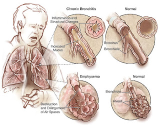
Management of COPD

Image
The goal of COPD management are: Touch ups the ability of people with symptoms mengatasiu not only in the acute phase, but also the chronic phase. Improving the ability of the patient in carrying out daily activities. Reducing the rate of progression of disease if the disease can be detected early. Management of COPD in the elderly are as follows: Etiological factors negate / precipitation, for example immediately stop smoking, avoid air pollution. Cleaning the bronchial secretions to help in various ways. Eradicate the infection with antimicrobial. In the absence of antimicrobial infections need not be given. Provision of appropriate antimicrobial should be in accordance with the germs that cause infections according to the results of sensitivity testing or empirical treatment. Overcome bronchospasm with bronchodilator drugs. The use of corticosteroids to resolve the inflammatory process (bronchospasm) is still controversial. Symptomatic treatment. Treatment of the complications tha...

Management of Occupational asthma

Image
To prevent the occurrence of occupational asthma is a medical examination prior to employment, use of protective equipment, monitoring air pollutants in the working environment is highly recommended. If occupational asthma has occurred, then transfer to the outside of the work environment is important. If for some reason can not be moved then it must be the prevention and monitoring of lung function decline. Periodic evaluation of lung function in workers who already suffer from occupational asthma are needed to prevent disability. Clinical asthma will persist until a few years even though the worker has been out of work environment. Medical treatment in patients with occupational asthma such as bronchial asthma in general: Theophylline , a bronchodilator and may suppress neutrophil chemotactic factor. Effectiveness of the two functions above depend on the serum levels of theophylline. Beta agonists , bronchodilators are the most good for the treatment of occupational asthma in com...

Management Of Pleural Effusion

Image
Management Of pleural Effution : Pleural fluid aspiration performed, to reduce the unpleasant taste or "discomfort" and shortness of breath.  It is advisable to gradually aspirations.Fluids released between 500 - 1000 cc. When taking too many and can quickly lead to pulmonary edema . Incorporating intrapleura chemotherapy for malignancy (eg radioactive gold or Ytrium). More often performed pleurodesis in the process of malignancy or in the frequent recurrence of effusion. By using 500 mg tetracycline powder is dissolved in 50 cc of physiologic saline. Patients shake it so flat, then the liquid removed after clamped for 24 hours or given jodium powder or talc. Pain that occurs because the administration of drugs above can be overcome with the analgesic. If need be given pethidine 100 mg i.m. Provision of steroids combined with antituberculosis can absorb the pleural effusion caused by pulmonary tuberculosis quickly and reduce fibrosis. Tags :  Pleural effution ,  Pleural...

Management of Chronic Bronchitis

Image
General: Education to patients and their families Stop smoking and avoid air pollution Prevent infection Clean environment Hydration moderation: drink enough water (8-10 glasses a day) Proper nutrition: protein rich diet and avoid heavy meals before bedtime, milk can cause increased bronchial secretions, should be prevented. Provision of drugs: 1. Bronchodilators: Aminophylline inj. 5.6 mg / kg i.v. or oral loading dose of 3 x 100-200 mg. Terbutaline 3 x 2.5 mg oral or injection 0.25 mg s.c. every 4-6 hours (1 mg / ml; = 2 ml ampoule). 3 x 2 mg salbutamol orally. 2. Expectorant: Water is a good expectorant Glyceryl guaiacolate 4 x 100-200 mg. 3. Mucolytic : Bromhexine HCL: 3 x 1 tablet by mouth. N-acetyl cysteine: 3 x 200 mg orally. 4. Respiratory therapy: Aerosols: Ipratropium Bromide 3 x 2 puffs Oxygen: 1-2 liters / minute via nasal cannula if PaO2 <55 mm Hg. 5. Rehabilitation: Physiotherapy Relaxation exercises Breathing exercises Chest percussion and postural drainage Exerci...

Management Of Hemothorax

Image
Hemothorax patient death can be caused because of the large blood loss and the occurrence of respiratory failure . Respiratory failure due to the large amount of blood in the pleural cavity pressure of lung tissue and reduced lung tissue that does ventilation. Then treatment hemothorax as follows: 1. Emptying of blood from the pleural cavity. Installed "chest tube" and is connected with the WSD system, this can accelerate the lung expands. 2. Stop the bleeding. If the installation of WSD, the blood still does not stop, then considered for thoracotomy. 3. General state of repair. Giving oxygen 2-4 liters / minute, the length adjusted to the clinical changes, better yet, if the monitored with blood gas analysis. Try to people with normal blood gases. Giving blood transfusion: seen from a decrease in Hb. As a benchmark can be used the following calculation, every 250 cc of blood (from patients with Hb 15 g%) can raise ¾ g% Hb. Given with a normal drop of about 20-30 drops / m...

Management Of Pneumothorax

Image
Outside the hospital. In light of spontaneous pneumothorax or pneumothorax simplex. Minimal or no complaints at all, are usually found by accident. The air in the pleural cavity will diresorbsi spontaneously. Because it does not require invasive measures. "Tension pneumothorax". Done in a sterile and carried out the stabbing in the sore area with a syringe the size of the largest. Stabbings in the space between the ribs into 2 in the front line of mid-clavicle. In young women (cosmetics) stabbings in the space between the ribs into 4 or 5 in the mid-axillary line. Then the needle tip covered with a sheet of thin rubber or thin plastic that can serve as a valve. Subsequently the patient was sent to hospital. I n the hospital. At the same place to do the installation of WSD, using trokar (troicar). It should be noted, that all actions undertaken SCARA sterile. WSD is removed, when the lung is expanding well and no complications after plastic hose clamped shut or 24 hours to pro...

Management of Empyema Thoracic

Image
The principle of treatment in empyema : 1. Emptying of the pleural cavity of pus 2. Antibiotics 3. Closure of the pleural cavity 4. Causal treatment 5. Additional treatment. 1. Emptying of the pleural cavity. a. Simple aspiration. Performed repeatedly using a large needle hole. This method is good enough to remove most of the pus or fluid from acute empyema is still runny. Losses such as these techniques often lead to "pocketed" empyema . Ultrasound can be used to determine the localization of "pocketed" empyema . b. Drainage is closed. Installation "= closed thoracostomy tube drainage (WSD)". Indications of this drain fitting, if the pus is very thick, pus is formed after 2 weeks and there has been piopneumotoraks. Installation of the hose should not be too low, the diaphragm is usually raised because of empyema . Select a hose that is large enough. If 3-4 weeks of no progress should be pursued by other means, such as in chronic empyema. c. Installation...

Management of Bronchiectasis

Image
A. Conservative: 1. Control of acute and chronic infection, mucus secretion, airway obstruction and complications, such as: coughing up blood, respiratory failure and cor pulmonale, in order to prolong life, improve quality of life and prevent disease progression. 2. Antibiotics if there is infection. 3. Chest physiotherapy and postural drainage with forced expiratory technique for removing secretions. 4. Aerosols with physiologic saline or beta agonists prior to chest physiotherapy may facilitate the release of sputum / secretions. 5. Bronchodilators to improve airflow, helping mukosilia clearance and physiotherapy improve outcomes. 6. Corticosteroids when there is severe bronchospasm ( CPOD or Asthma Bronchiale ). B. Surgery: Indications of surgery: Local Bronchiectasis Haemoptoe massive. Tags : bronchiectasis pneumonia , what is bronchiectasis , bronchiectasis copd , bronchiectasis symptoms , bronchiectasis treatment , bronchiectasis definition , cystic bronchiectasis , Management b...

Management of Lung abscess

Image
1. General management: Improving the general condition of patients with high-calorie high-protein diet and drink plenty of fluids. a. Antibiotics. Procaine penicillin G given 1.2 million units im every 12 hours + chloramphenicol 500 mg every 6 hours for 10-15 days. or Procaine penicillin G 1.2 million units i.m. every 12 hours + Metronidazole 500 mg every 6 hours for 10-15 days. or Clindamycin 600 mg every 8 hours for 10-15 days. b. Postural drainage and physiotherapy. The position of the body are arranged so that pus can come out by itself (due to gravity) or with the help of the physiotherapist. 2. Special Treatment: a. Bronchoscopy If pus is difficult exit, it is necessary to bronchoscopy to clear the airway and sucking pussy. b. Surgery When chemotherapy failed. A chronic abscess, cavity remains and sputum production remained there while the clinical symptoms are still present after adequate therapy for 6 weeks or the rest of extensive scar tissue that can interfere with l...

Management of Bacterial Pneumonia

Image
Indications of hospitalization is: 1. Patients with basic diseases 2. Patients with complications. General management. 1. Correction of underlying abnormalities (underlying disease) 2. Bedrest 3. Symptomatic medications, administered only if necessary, such as: 3 x 500 mg Paracetamol (in hyperpyrexia) Morphine 10 mg s.c. (If there is pain severe). 4. Maintain fluid and electrolyte balance with the help of intravenous fluids, 5% dextrose, normal saline or Ringer's lactate. 5. Selection of anti-infective drugs. Selection of antimicrobial drugs, should be based on sensitivity tests and sensitivity, but due to time and facilities greatly affect the success of this test, then giving more medicine based on empirical, as below: Special Treatment: 1. Pneumococcal pneumonia, Basic disease : elderly,  Chronic Obstructive Pulmonary Disease (COPD) , CHD, diabetes mellitus, alcoholism, post-influenza. Clinical features : sudden illness, high fever, chills, pleuritic pain, cough productive ...

Management Bronchial Asthma

Image
Procedure Of Bronchial Asthma Non-pharmacotherapy Provision of O2 Fluid Postural Drainage Avoiding exposure to allergens Guidance on patients and families about asthma, causes, and how to overcome them. Avoid the trigger factors (diet, medication, living habits, allergens) Immunotherapy / desentisisasi Physiotherapy breath, vibration and / or thoracic percussion, an efficient cough. Pharmacotherapy: 1. Bronchodilators: - Adrenaline; solution of adrenaline 1: 1000 subcutaneous 0.3 cc waiting for 15 minutes, if it has not subsided again given 0.3 cc if it has not abated, can be repeated once again 15 minutes later 0.3 cc. For children can be given smaller doses: 0.1 to 0.2 cc. Caution in elderly patients (coronair heart disease), hypertension, hyperthyroidism. -  Beta 2 agonists (oral, injection, inhalation / MDI, nebulizer) Orsiprenalin (alupent) 3 x 20 mg orally; subcutan 3 x 0.25 mg; 3 x 3 spray inhalation. Bricasma subcutan 3 x 0.5 cc. Heksoprenalin (Ipradol) 3 x 0.50 mg orally. ...