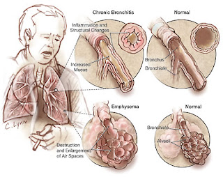
Posts

Showing posts from August, 2011

Bronchopneumonia

Image
Bronchopneumonia   is a type of lung infection caused by infectious agents and are in the area around the bronchi and alveoli. Etiology In general, individuals who are stricken with bronchopneumonia caused by a decrease in the body's defense mechanism against the virulence of pathogenic organisms. People who have a normal and healthy body's defense mechanisms against respiratory organs which comprises: glottis and the cough reflex, presence of mucous layer, the movement of cilia that move the bacteria out of the organ, and local humoral secretion. Incidence of bronchopneumonia caused by viruses, bacteria, fungi, protozoa, mycobacteria, mycoplasma, and rickettsial. , among others: Bacteria: Streptococcus, Staphylococcus, H. Influenzae, Klebsiella. Virus: Legionella pneumoniae Fungi: Aspergillus species, Candida albicans Aspiration of food, oropharyngeal secretions or gastric contents into the lungs Pulmonary congestion occurs because the old one. Another cause of pneumonia is...

Pleural Effusion

Image
Pleural Effusion is a collection of fluid in the pleural cavity or cavity between the parietal pleura and visceral pleura, can be fluid transudates or exudates. Pathophysiology and Etiology - Transudates: An extracellular fluid that collects in the pleural lavum passively. With a specific gravity of the liquid is less than 1.015; protein in the fluid of less than 2-3 g / dl. Can occur as part of a general or edema in left heart failure. Generally the cause is: an increase in pulmonary venous pressure, hypoalbuminaemia and mediastinitis fibrosis or miksedema. - Exudate: Fluid in the pleural cavity caused by infectious diseases or neoplasms, generally protein levels> 3 g / dl, yellow or orange, with or without cells or bacteria. Generally it can be caused by inflammation, neoplasm, and abnormal lymph drainage. Clinical Symptoms Shortness of breath is the main symptom, sometimes accompanied by feelings of discomfort in the chest. When pleural fluid is low, can not be detected by clin...

Management Of Pleural Effusion

Image
Management Of pleural Effution : Pleural fluid aspiration performed, to reduce the unpleasant taste or "discomfort" and shortness of breath.  It is advisable to gradually aspirations.Fluids released between 500 - 1000 cc. When taking too many and can quickly lead to pulmonary edema . Incorporating intrapleura chemotherapy for malignancy (eg radioactive gold or Ytrium). More often performed pleurodesis in the process of malignancy or in the frequent recurrence of effusion. By using 500 mg tetracycline powder is dissolved in 50 cc of physiologic saline. Patients shake it so flat, then the liquid removed after clamped for 24 hours or given jodium powder or talc. Pain that occurs because the administration of drugs above can be overcome with the analgesic. If need be given pethidine 100 mg i.m. Provision of steroids combined with antituberculosis can absorb the pleural effusion caused by pulmonary tuberculosis quickly and reduce fibrosis. Tags :  Pleural effution ,  Pleural...

Bronchial Asthma

Image
Bronchial asthma  is a disease of the lung inflammation in the airways resulting in airway hiperrespon to various stimuli that can cause constriction of the airways that can arise thorough so shortness of breath that is reversible either spontaneously or with therapy. Etiology Genetic factors Environmental factors Materials allergens Respiratory tract infections (especially viruses) Air pollution Food Factor Trigger factors Allergens Physical Chemicals Infection Mechanical factors Psychological factors Pathogenesis The entry of allergens into the airway will cause a reaction between the allergen with immunoglobulin E. The release of materials from mastosit mediators, which cause the inflammation in the bronchial mucosa and submucosa causing bronchial smooth muscle contraction. Pathology Infiltration of inflammatory cells such as eosinophils, neutrophils, and other airway epithelial damage, resulting in expenditures as well as mediators and thickening of the mucosal and submucosal e...

Emphysema

Image
Emphysema Lung Is the permanent widening of the structure of pulmonary gas exchange that is distal to the terminal bronchioles, accompanied by destruction of alveolar walls. Etiology Main factors: smokers either active smoking or passive smoking depending on the length, number, how to smoke. Air pollution, in the form: Particles, These chemicals, Toxic Gas Infections: viral and bacterial Genetics: a. Talent arise emphysema b. The balance of destructive enzymes (protease) & protective enzymes (alpha-1-antitrypsin). Google Image Pathogenesis Inhalation of cigarette smoke or pollutants stimulate cells in the lung macrophages and neutrophils produce elastase and collagenase is an enzyme that damages the fibers of elastin and collagen, and which is the framework of the alveoli and acini in order not to collapse. Smoking also inhibit the action of alpha-1-antitrypsin, an enzyme that protects elastin fibers against proteases. In the lung there is a balance between destroyi...

Special examination of Chronic bronchitis

Image
Special examination :  X-ray photo PA and left lateral chest -  Typically show no abnormalities, except when it occurs cor pulmonale or emphysema. -  It may also seem an increase in "bronchovascular markings". Sputum: direct smear, Gram staining. Please note: the existence of cells in sputum, whether the material is indeed contaminated with sputum or saliva from the oropharynx. Sputum can be identified because of alveoler macrophages (histiocytes). Macrophages alveoler this form round cells, large, with eccentric nuclei and oval or kidney-shaped near the edge of the cell. Tues skuamus flat and very large and has a spherical nucleus in the middle. If there were> 25 epithelial cells in sputum skuamus, means contaminated with saliva or secretions from the oropharynx. In chronic bronchitis who experience eksaserbrasi yellow sputum, bronchial ciliated epithelium that despite apparent in sputum neutrophil and looks. At the time of remission decreased number of neutrophils, b...

Chronic Bronchitis

Image
Chronic bronchitis is a disease, characterized by cough with phlegm every day, at least 3 months per year and lasts for 2 consecutive years, and the absence of other diseases with symptoms. Etiology and pathogenesis Etiology Exposure to cigarette smoke, both at the "smoker" or "passive smoker". For the smoker is usually measured by pack year (pack years). Exposure to contaminated air in workplaces (eg dust or gas from industrial or workplace). Social factors and residential solid and air pollution in the settlement area by the gases SO2 and NO2. Respiratory tract infection by the virus will cause eksaserbrasi chronic bronchitis. For example, by class sinsisial respiratory viruses and influenza virus group. Most bacteria that are found in chronic bronchitis are streptococcus pneumonia and haemophilus influenza. Pathogenesis a. Cigarette smoke is a disincentive to the bronchial mucosa, to changes: Lung Defense: Purge function mukosilia slow, Alveoler Macrophage numbe...

Management of Chronic Bronchitis

Image
General: Education to patients and their families Stop smoking and avoid air pollution Prevent infection Clean environment Hydration moderation: drink enough water (8-10 glasses a day) Proper nutrition: protein rich diet and avoid heavy meals before bedtime, milk can cause increased bronchial secretions, should be prevented. Provision of drugs: 1. Bronchodilators: Aminophylline inj. 5.6 mg / kg i.v. or oral loading dose of 3 x 100-200 mg. Terbutaline 3 x 2.5 mg oral or injection 0.25 mg s.c. every 4-6 hours (1 mg / ml; = 2 ml ampoule). 3 x 2 mg salbutamol orally. 2. Expectorant: Water is a good expectorant Glyceryl guaiacolate 4 x 100-200 mg. 3. Mucolytic : Bromhexine HCL: 3 x 1 tablet by mouth. N-acetyl cysteine: 3 x 200 mg orally. 4. Respiratory therapy: Aerosols: Ipratropium Bromide 3 x 2 puffs Oxygen: 1-2 liters / minute via nasal cannula if PaO2 <55 mm Hg. 5. Rehabilitation: Physiotherapy Relaxation exercises Breathing exercises Chest percussion and postural drainage Exerci...